– Colleen Rollins, 24th January, 2020
PhD student, Department of Psychiatry, University of Cambridge; Gates Cambridge Scholar researching how the structure and function of the brain supports the experience of hallucinations
The world you perceive is not through passive recording of your environment, like a camera capturing film. Rather, it is coloured by past experiences, by how things have previously looked and sounded, by beliefs and emotions. A compelling example of this is illustrated by visual illusions, such as the Ponzo illusion (Image 1), in which our perception of a scene does not match the visual input we receive. Even though the two horizontal lines in the image are identical in length, we experience the farther line as longer. But if perception doesn’t accurately map to physical reality, how do we know which of our sensory experiences are real?

Image 1. The Ponzo illusion
One hypothesis is that there are decision making processes involved in distinguishing between internally-generated information and information present in the outside world. This attribution process is called “reality monitoring”. There is evidence that people with a diagnosis of schizophrenia are more likely to make errors on reality monitoring tasks, and that patients who experience hallucinations are even more likely to make errors than those who do not. The question my research aims to answer is how are these anomalous perceptions – hallucinations – are supported by the brain.
During the third trimester of pregnancy, the developing brain undergoes a transformative process known as “cortical folding” or “gyrification”, in which it matures from a smooth surface to one that is highly convoluted. The peaks of the brain’s folds are called “gyri”, and the troughs, “sulci” (Image 2). Once established, the pattern of folding is generally stable across a person’s lifespan and is unique like a fingerprint. What’s more, the organization of folds is intrinsically related to large scale resting-state networks (RSN) – systems of interacting brain regions that show common patterns of functional activity when an individual is engaged with specific cognitive tasks, such as attention or language tasks. Some RSNs may already be established in early childhood, and perhaps even before birth. This includes the salience network, which engages brain regions involved in detecting the prominence of internal and external stimuli, and has been robustly associated with hallucinations in schizophrenia. Not to be forgotten is that the brain networks supporting hallucinations must also be sensitive to life experiences, since trauma is a strong risk factor for hallucinations, and culture can affect the content and meaning of hallucinations.
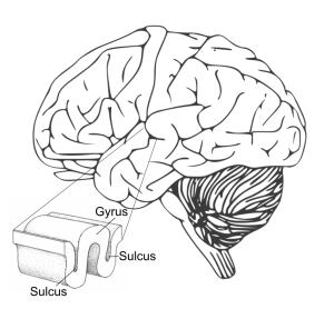
Image 2. Schematic of an adult human brain. The brain is folded in a complex pattern of “hills”, called gyri, and “valleys”, called sulci.
Recent research at the University of Cambridge has shown significant associations between reality monitoring, hallucinations in schizophrenia, and brain folding patterns in the medial prefrontal cortex (mPFC), an area implicated in attending to self-referential information. Specifically, absence of the paracingulate sulcus (PCS) on both sides of the brain, a fold in the mPFC, has been associated with reduced reality monitoring ability, and reduced length of the PCS with hallucination occurrence in people with a diagnosis of schizophrenia.
Our current study hopes to leverage two ethnically independent structural magnetic resonance imaging (MRI) datasets of schizophrenia patients to empirically test theoretical predictions linking sulcal topology, reality monitoring and salience and auditory network integrity. We re-purposed data from two independent studies recruiting patients with schizophrenia who underwent clinical assessment and structural MRI. The datasets included a predominantly White British sample assessed in multiple sites across the United Kindgom, and a Han Chinese sample assessed in Shanghai, China. Patients were grouped into those with and without hallucinations based on their score on a clinical scale that assesses hallucination occurrence and severity. Healthy controls who did not experience hallucinations were additionally recruited in the Shanghai sample. Datasets were matched with respect to sex, age, and hallucination severity.
Our first main result was that the length of the left PCS was reduced in patients with hallucinations, as has been shown previously, in both ethnically distinct datasets a finding that remains significant when controling for other factors that may influence sulcal length, such as age, sex, scanner site, and total intracranial volume. A major limitation of measuring the PCS is that it shows high intersubject and interhemispheric variability – it may be present, absent, fragmented, or intersected by other sulci. Therefore, no fully automated method exists to measure the PCS. A major part of the present study has been developing and assessing methodologies to automatically quantify the PCS. This has involved manually tracing the PCS, comparing it to semi-automated methods, and partnering with the Computer Science and Technology Department at the University of Cambridge to create a machine learning model to automatically detect the PCS from a novel MRI scan.
We next aimed to link this specific sulcal anomaly to the broader context of neurodevelopment, using structural covariance networks (SCN), which reflect developmental coordination between brain regions. We calculated the local gyrification index, a measure of the degree to which the brain is folded, for each brain region delineated by the Human Connectome Project multimodal atlas (a map of the human brain). We merged brain regions based on their belonging to one of eight established large-scale RSNs. Comparing SCNs between groups, we found increased gyral synchrony between auditory and salience networks, suggesting that alterations before or shortly after birth to the structural integrity of developing salience and auditory networks may increase risk of experiencing hallucinations.
Like many research findings, this result raises more questions than it answers. Are sulcal anomalies specific to schizophrenia, or do they present in other disorders in which hallucinations manifest, such as borderline personality disorder or Parkinson’s disease? How do neurodevelopmental events like cortical folding blueprint or mediate subsequent life experiences? What are the mechanisms driving cortical folding and how do they affect brain function?
With this study, we identified a structural network of sulcal deviations and increased gyrification covariance between salience and auditory networks associated with the experience of hallucinations, across two ethnically independent datasets. The impact of this research is two-fold: On the one hand, there is hope that a better understanding of the brain processes underlying hallucinations will lead to more effective and targeted treatments. On the other, hallucinations offer an interesting window into fundamental questions on the nature of perception, the boundaries between self and other, internal and external, and how we construct our reality (Image 3).

Image 3. Scatterbrain. Acrylic and Sharpie Pen on A0 Card. Artwork by sk172, an artist who has been experiencing florid psychotic symptoms for over a decade, which he treats with clozapine. His artwork is a dialogue between internal stimuli and external stressors, with a unique style merging urban-street narrative and primitivism drawing. @sk_oneseventwo
